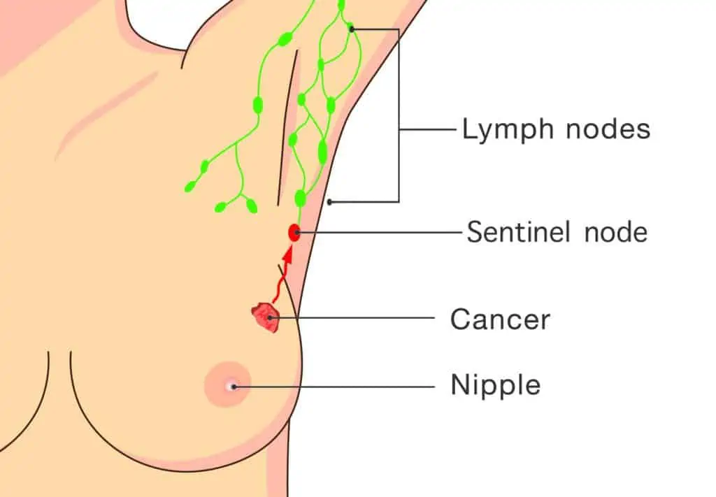
The sentinel lymph node(s) are the first lymph nodes that the breast lymphatics drain into. If cancer cells have spread from a breast cancer, it is likely that they will travel through the sentinel node before moving through to other lymph nodes. The most common location for a sentinel lymph node to be located is in the armpit (axillary nodes) on the same side of the affected breast. Less commonly, they are found in the lymph nodes between the ribs near the sternum (internal mammary nodes), above the collar bone (supraclavicular nodes), and rarely, in the axilla on the other side.
Prior to the introduction of sentinel node biopsies in 1991, an axillary clearance (or axillary dissection) was the standard approach to assessing the lymph nodes in breast cancer surgery. This meant that many lymph nodes were removed which carries the associated risks of greater discomfort, numbness, shoulder stiffness and potential lymphoedema. Modern studies have proven the safety and effectiveness of sentinel node biopsy which reduces the need for an axillary clearance, and the associated side effects.
What is a sentinel lymph node biopsy?
A sentinel lymph node biopsy identifies those important first draining lymph nodes so that they can be removed and tested by a pathologist to detect if cancer has spread to them. Studies have shown that up to 30% of women with early breast cancer will have cancer cells in their sentinel lymph nodes.
From here, decisions can be made about whether more surgery to the axilla is required. It also helps to provide information about prognosis, and whether you will be recommended other adjuvant treatments (for example, radiotherapy, endocrine therapy or chemotherapy).
How do you find the sentinel lymph nodes?
There are a few techniques that surgeons use to find the sentinel node. These include lymphoscintigraphy and Patent V Blue Dye.
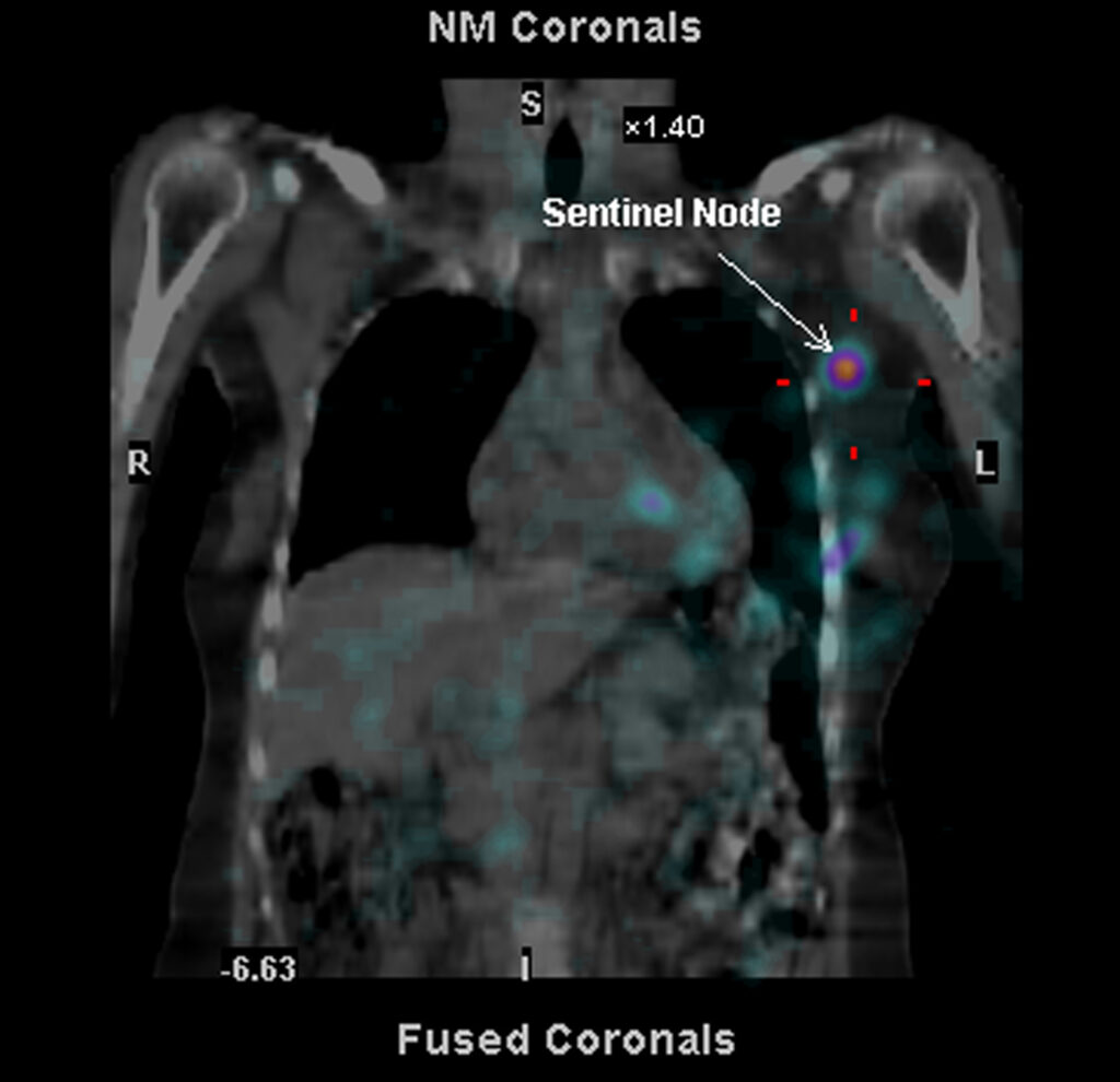
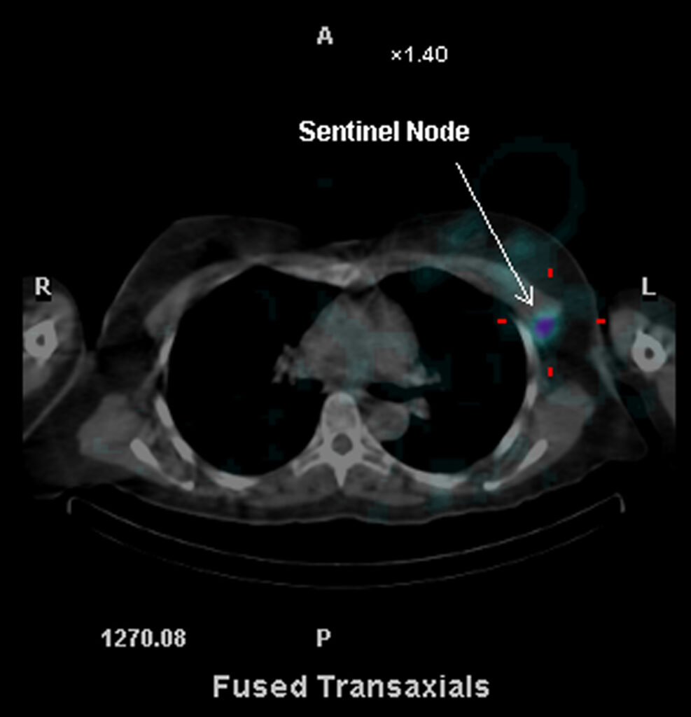
Lymphoscintigraphy involves a radiologist injecting a radiolabelled tracer into the breast the day before or the morning of your surgery. A special scan is done which helps the surgeon identify the location and number of sentinel lymph nodes (see images below). A ‘gamma probe’, which makes a sound when it is close to the sentinel nodes, is used to locate them during the operation.
Blue dye may also be injected into the breast when you are asleep at the time of your operation. It will temporarily stain the lymph nodes bright blue to help highlight them during the operation, making them easier to see and find.
Unfortunately, a sentinel node biopsy is not suitable for everyone. Dr Lancashire will discuss whether a sentinel node biopsy is appropriate for you.